
Case Study - The Bruise That Solved the Mystery
How a routine blood draw was mistaken for a surgical complication — and what the anatomy actually shows
by Shanise Keith • July 28, 2026
A woman goes into surgery for a craniectomy to treat trigeminal neuralgia — a procedure with nothing to do with her arm. Partway through, the surgical team monitoring her nerve signals notices something: the readings from her left arm are dropping. Their first assumption is the obvious one. She’s been positioned on the table for hours, and pressure on the brachial plexus from positioning is a known risk during long procedures. They adjust her arm. The signal partially recovers. Surgery continues.
She wakes up unable to fully feel or move part of her hand.
The team does exactly what you’d expect a good team to do — they investigate the surgery. They review the positioning notes. They check the padding, the table, the anesthesia record, everything about how she was lying during a multi-hour procedure. All of it points toward a positioning-related brachial plexus injury, the kind that’s a recognized, if uncommon, risk of long surgeries.
But that’s not what happened to her.

Someone eventually notices bruising at her elbow crease — from a routine blood draw done before she ever went into the operating room. Nerve conduction studies confirm it: this wasn’t a brachial plexus injury from surgical positioning at all. It was a median nerve injury from the venipuncture itself, hours before the surgery even started. The case report doesn’t identify which vein was used or exactly what caused the bruising and compression — only that bruising was present at the site and that a compression-type injury from bleeding under the skin is a well-documented mechanism for this kind of nerve injury.
That’s also a plausible reason she didn’t notice anything wrong right away: that kind of compression builds gradually as blood collects under the skin, so a deficit from it might not show up the moment the needle goes in — it can take time to press on the nerve hard enough to cause a problem, time she happened to spend under anesthesia and unable to feel it happening. The dramatic, complicated explanation was wrong. The routine blood draw was right.
What a “nerve injury” actually feels like
Let’s pause on what we’re actually talking about, because “nerve injury” sounds abstract until you connect it to something everyone has felt.
You know the feeling of your arm falling asleep — you slept on it wrong, or you had it bent under you too long, and it goes numb and tingly until you shake it out and the feeling comes back in a minute or two. That’s a nerve being compressed temporarily, and it recovers on its own because nothing was actually damaged.
A real nerve injury can start out feeling similar, but the sensations it produces can go well beyond numbness and tingling. Depending on which nerve fibers are affected and how severely, patients describe shooting or electrical pains that travel down the arm, or a burning sensation that doesn’t let up. Some describe it as feeling like the sensation you get when you hit your “funny bone,” except it doesn’t fade — it lingers, or comes back with movement. The type and severity of the injury shapes which of these a patient experiences: a mild compression might produce lingering numbness, while direct trauma to the nerve can produce that sharp, electrical, shooting quality that patients often describe as far more alarming than ordinary numbness.

A venipuncture-related nerve injury is that same mechanism, but with the potential to not just go away. It can happen a few ways:
- Direct trauma — the needle itself nicks, punctures, slices, or severs the nerve on the way to (or instead of) the vein.
- Compression from a hematoma — bleeding under the skin after the draw forms a pocket of blood that presses on the nerve, the same way your body weight compressed your arm while you slept, except this pressure doesn’t go away when you shift positions. It stays until the hematoma resolves, or sometimes longer.
- Stretch or positioning — less common, and not venipuncture-related, but relevant to this post. This can happen in the operating room, or with an unconscious person, where a limb can be held in one position for hours.
The difference between “my arm’s asleep for a minute” and a real nerve injury is duration and severity of pressure. A nerve that’s compressed briefly recovers fully. A nerve that’s compressed hard enough, or long enough — from a needle, from a hematoma, from prolonged positioning — can end up with lasting numbness, weakness, or burning pain that doesn’t resolve on its own. That’s the difference between an inconvenience and an injury that needs medical follow-up.
Why this keeps happening
This isn’t as rare as it sounds. In one study (linked at the bottom of the article), a review of iatrogenic (meaning we caused it) nerve injury claims accepted through a no-fault compensation system found venipuncture ranked as the second-most common documented cause of these injuries in that dataset — behind surgical positioning — with the median nerve most often affected. That’s a reflection of what showed up in accepted compensation claims, not a statement about how often either cause occurs across all surgical and medical care generally. Positioning-related nerve injuries are themselves uncommon overall, even though they show up disproportionately often in claims data. Still, the fact that venipuncture ranked where it did in a real claims review means this isn’t a freak occurrence tucked away in a single case report. It’s a documented, recurring pattern.
The anatomy explains why. At the elbow crease, the median nerve typically runs close to the brachial artery — one ultrasound-based study measured the gap at roughly 1.4 millimeters, the kind of proximity that doesn’t leave much margin for error. A larger ultrasound review of 68 patients found that closeness isn’t a fixed number, though: the separation ranged from as little as 2.5mm to as much as 26mm (about 1 inch), with about 1 in 10 patients showing more than 12.5mm (about half an inch) of space between the two. What did stay constant was direction — in every one of those 68 patients, the nerve sat medial to the artery, even as the distance between them swung widely.
Textbook diagrams show a fairly standard layout: cephalic vein toward the thumb side, basilic vein toward the pinky side, artery and nerve running together medially near the basilic vein. But the basilic vein’s relationship to the artery isn’t fixed the way the nerve’s is — sometimes the artery sits medial to the vein, sometimes lateral, sometimes almost directly underneath it. So while the nerve is typically on the medial side of the artery, just at a variable distance, the vein you’re actually aiming for can be on either side of it entirely. A vein that looks like it’s sitting in a “safer” spot on one patient might be sitting right on top of the danger zone on the next.
The point is, we don’t know exactly where the nerve is, even when we can pinpoint the artery. It’s usually medial, but everyone’s anatomy is different — it could potentially sit lateral instead. That’s exactly why the medial aspect of the antecubital fossa is to be avoided. There’s no knowing precisely where the nerve sits on a given patient. We just know there’s a nerve here somewhere.
Why the rule doesn’t bend
This is exactly why CLSI’s guidance isn’t “measure a safe distance” or “palpate the artery and use your judgment.” It’s a categorical instruction: CLSI’s PRE02 standard states that veins in the medial aspect of the antecubital area should be off the table entirely unless no other vein offers a confident, safe, successful option for collection.
It would be reasonable to ask why the standard doesn’t just say “stay a finger-width away from the artery” or “palpate first, then decide.” The answer is the anatomy itself. If everyone’s vessels and nerves lined up with the textbook every time, a measured distance or a palpation check might be enough. But because that relationship is genuinely unpredictable from patient to patient, a rule that depends on real-time judgment in a variable landscape isn’t a reliable safeguard. A hard boundary is. CLSI isn’t asking phlebotomists to get better at estimating distances near a nerve and artery — it’s removing the estimate from the equation entirely by taking that whole region off the table when there’s a viable alternative.
That’s also why the case above deserves more than a passing mention as a surgical curiosity. There isn’t enough detail in the report to know what, if anything, went wrong in how the draw was performed. What we do know is that it was a routine draw, done the way thousands are done every day, and the injury happened anyway because of where the needle went, and how the hematoma developed. That’s precisely the scenario a categorical rule is designed to prevent: not the reckless outlier, but a draw that went wrong.
The detail that solved it
What eventually cracked the case wasn’t advanced testing — it was someone noticing a bruise at an elbow crease and asking a question instead of assuming it was unrelated to what was happening in the operating room. The nerve conduction study confirmed what the bruise suggested. But the bruise is what made someone look in the right place at all.
That’s a good habit to carry into daily practice, well beyond this one case: when a patient reports something odd after a draw — tingling, burning, a sensation that doesn’t feel like a normal stick — that report deserves attention, not reassurance that it’s probably nothing. Sometimes it’s the only clue anyone gets.
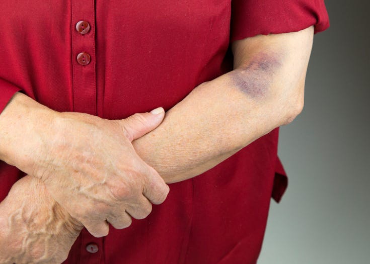
I don’t know for certain which vein was used in this case — the report doesn’t say, and neither does it say exactly what caused the bruising underneath. But given everything laid out above about where the basilic vein tends to sit relative to the median nerve and brachial artery, if I had to put money on it, that’s where I’d put it. It fits the pattern too well not to suspect it: a vein in that medial territory, in a patient whose anatomy put the nerve close enough for a routine stick to end in a compression injury. I can’t prove that’s what happened here. But it’s exactly the scenario the rest of this piece has been building toward.
But the better goal is never needing that clue in the first place. The median cubital and cephalic veins should be the default choice whenever they’re available, precisely because they sit farther from this neurovascular territory. The basilic vein sits close enough to the artery and nerve, on enough patients, that it should be avoided whenever another option exists — not reached for as a routine alternative. Treating it as a last resort, rather than an equal option on the list, is what actually prevents cases like this one from happening at all.
Related Posts and Information
overall rating: my rating: log in to rate
case study hematoma basilic brachial nerve injury pain