
The Order of Draw Series: Part Four - Heparin Tubes
This week, we're examining green top heparin tubes—and discovering why tubes that prevent clotting must come after tubes that depend on clotting.
by Shanise Keith • January 27, 2026
Why Green Tops Come Fourth
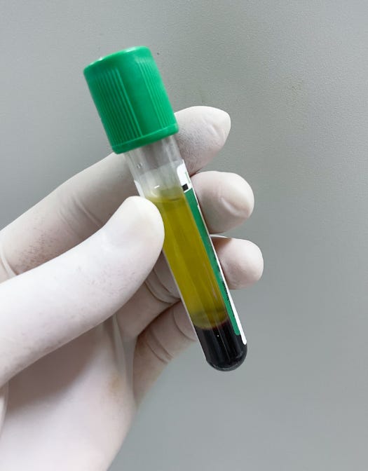
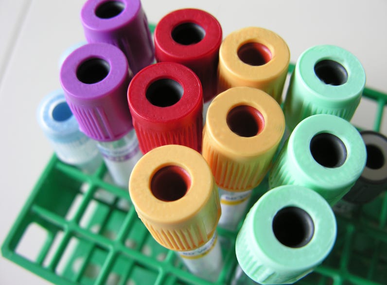
After blood cultures, light blue coagulation tubes, and serum tubes, the order of draw brings us to green top tubes containing heparin. These tubes occupy the fourth position for a critical reason: they must come AFTER serum tubes (to avoid preventing the clotting that serum collection requires) but BEFORE EDTA tubes (to prevent cross-contamination that affects both chemistry and hematology results).
Contamination can happen in two ways when green tops are drawn out of sequence:
If green tops are drawn before light blue citrate tubes: Heparin residue on the needle can transfer into the citrate tube, potentially causing falsely prolonged coagulation times. Even trace amounts of heparin dramatically affect coagulation testing—the aPTT is particularly sensitive and can be markedly prolonged, while the PT will also be affected. A patient with normal coagulation could appear to have a bleeding disorder, leading to delayed emergency surgeries, unnecessary hematology workups, and clinical decisions based on artifactual results. While heparin contamination more commonly occurs from drawing blood through heparinized IV lines, maintaining proper order of draw prevents any possibility of needle carryover contamination. This is why light blue citrate tubes must come before green heparin tubes.
If green tops are drawn before serum tubes: Heparin residue on the needle transfers into serum tubes, preventing the blood from clotting properly. The result is specimens with fibrin strands floating in partially clotted serum—unusable for testing.
If EDTA tubes are drawn before green tops: EDTA residue on the needle transfers into the heparin tube, creating a chemistry nightmare. EDTA chelates calcium and magnesium (making both appear falsely low), while the potassium in K2EDTA falsely elevates potassium levels. A patient with normal electrolytes suddenly appears to have life-threatening abnormalities requiring emergency treatment they don’t actually need.
By placing green tops after serum tubes but before EDTA tubes, we prevent both problems: serum tubes get clean blood that can clot properly, and heparin tubes get clean blood without calcium-chelating EDTA contamination
Types of Heparin: Not All Green Tops Are Equal
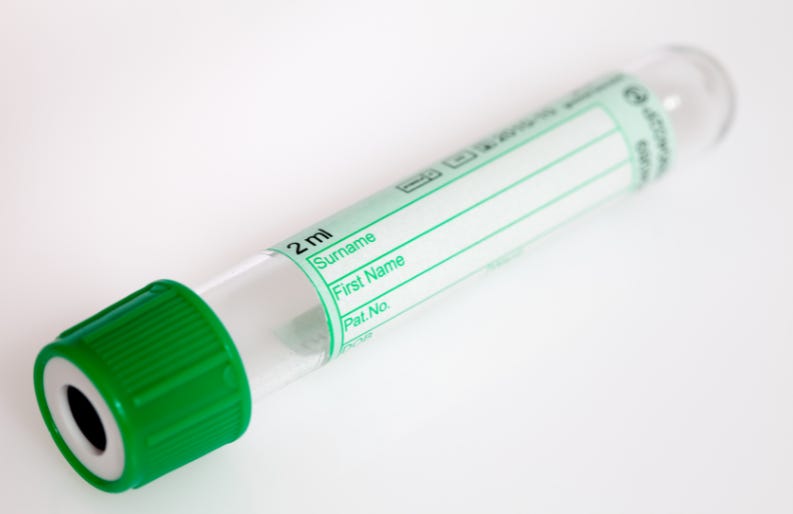
Here’s where heparin tubes get interesting: the tube cap might be green, but what’s inside varies. Different salt forms of heparin are used for different testing purposes, and understanding these differences matters for accurate results.
Lithium heparin (light green cap) is the most common. It’s used for routine plasma chemistry testing—electrolytes, kidney function, liver function, and most other chemistry panels. Lithium heparin became the standard because lithium doesn’t interfere with most chemistry assays. However, if you’re specifically testing lithium levels (for patients on lithium therapy for bipolar disorder for example), you obviously can’t use a lithium heparin tube—the tube additive would falsely elevate the result.
Sodium heparin (dark green cap) is used when you need to avoid lithium contamination, most commonly for lithium level testing. It can also be preferred for certain molecular biology applications and chromosome studies where lithium might interfere with cell culture.
Ammonium heparin is rarely used today but exists for specialized testing where both sodium and lithium could cause interference.
PST tubes (Plasma Separator Tubes) contain lithium heparin plus a gel separator, similar to how SST tubes contain clot activator plus gel. The gel creates a barrier between plasma and cells after centrifugation, making specimen handling easier and reducing the risk of cellular contamination. PST tubes are typically light green with a different shade or marking to distinguish them from plain lithium heparin tubes.
The salt form matters because it becomes part of the plasma specimen. If you’re measuring sodium levels and use a sodium heparin tube, or measuring lithium levels with a lithium heparin tube, you’ll get falsely elevated results from the tube additive itself.
*Note: While the colors listed above are typically what you may commonly see, different brands use different shades of green for their various heparin tubes. It’s very important to read the label to be sure you have the correct version, don’t trust the color.
The Discovery of Heparin: From Liver to Laboratory
The story of heparin begins in 1916 at Johns Hopkins University, where a medical student named Jay McLean was studying the clotting properties of various tissue extracts. He was working in the laboratory of physiologist William Howell, investigating procoagulant substances—things that made blood clot faster.
Instead, McLean discovered the opposite: an extract from liver tissue that prevented blood from clotting. Howell continued the work and named the substance “heparin” from the Greek word “hepar,” meaning liver. For years, scientists thought heparin was unique to liver tissue, though we now know it’s found in many tissues, particularly in mast cells and basophils.
The path from discovery to medical use took decades. Heparin’s chemical structure proved incredibly complex—it’s a large, heterogeneous mixture of polysaccharide chains, not a single molecule. Purifying it for medical use and determining safe, effective doses required extensive research. By the 1930s, heparin was being tested for preventing blood clots during surgery. By the 1940s, it became a standard anticoagulant for treating and preventing thrombosis.
The transition to blood collection tubes came later, as laboratories recognized heparin’s advantages for certain types of testing. Unlike anticoagulants that chelate calcium (EDTA, citrate), heparin doesn’t remove calcium from the specimen, making it suitable for tests that measure calcium and other electrolytes. And because heparin works immediately—no waiting 30 minutes for clotting—plasma from heparin tubes could be tested faster than serum, making it ideal for stat chemistry panels.
How Heparin Works: A Different Approach to Anticoagulation
Understanding why green tops must come after serum tubes requires understanding how heparin prevents clotting—and why its mechanism is so different from other anticoagulants.
Heparin doesn’t remove anything from blood. Unlike EDTA, which chelates calcium and physically binds and blocks it from the solution, heparin works by dramatically amplifying the activity of antithrombin III, a naturally occurring protein in blood that inhibits clotting factors.
Think of antithrombin III as a slow-acting security guard trying to stop several intruders (thrombin, Factor Xa, and other clotting factors). The guard can catch them eventually, but it takes time. Heparin is like giving that guard superpowers—suddenly the guard becomes 1,000 times faster and more effective. Antithrombin III normally inhibits thrombin at a relatively slow rate. When heparin binds to antithrombin III, it causes a conformational change that makes antithrombin III bind to thrombin almost instantaneously.
The effect is immediate and profound. Thrombin is the enzyme that converts fibrinogen to fibrin—the final step in clot formation. By inhibiting thrombin, heparin stops clots from forming. But heparin also inhibits Factor Xa and several other clotting factors, blocking the coagulation cascade at multiple points.
This is why heparin contamination in serum tubes is so problematic. Remember, serum tubes need blood to clot. They contain clot activators (silica or thrombin) specifically designed to trigger clotting. If heparin gets into that tube—even in trace amounts—it inhibits the very thrombin that’s supposed to be converting fibrinogen to fibrin. You get incomplete clotting: a weak, partial clot with fibrin strands that don’t fully separate from the serum. The specimen becomes unsuitable for testing.
This is why order matters. Green tops must come after red/gold serum tubes to prevent heparin from contaminating tubes that need to clot.
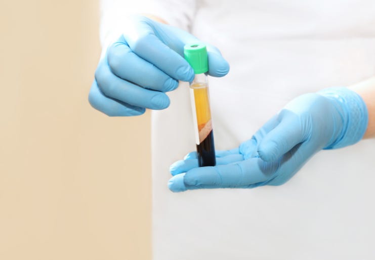
Plasma vs. Serum: Why It Matters
Heparin tubes produce plasma, not serum. This distinction is important for understanding their place in the order of draw and their uses in laboratory testing.
Plasma is the liquid portion of blood that’s been anticoagulated—all the clotting factors are still present, but they’ve been prevented from doing their job. Serum is what you get after blood clots—the clotting factors have been consumed in the clotting process, and what remains is plasma minus fibrinogen and several other clotting proteins.
For most chemistry testing, plasma and serum are interchangeable. Electrolytes, kidney function tests, liver enzymes, lipids—these analytes are present in both plasma and serum at essentially identical concentrations. But there are important differences:
Speed: Plasma from heparin tubes is available immediately after centrifugation. No waiting 30 minutes for clotting. This makes heparin tubes ideal for stat chemistry panels in emergency departments when physicians need results quickly.
Specimen quality: Because there’s no clotting process, there’s no clot retraction, no platelet activation, and no release of cellular contents during clot formation. This can reduce some sources of specimen variation.
Volume: You get slightly more plasma from a tube than serum from the same volume of blood, because no blood is trapped in a clot.
Test compatibility: Some tests specifically require serum (because clotting factors interfere) or specifically require plasma (because the test measures something consumed during clotting). Most chemistry tests work with either, but you need to know your laboratory’s validated specimen types.
What Tests Use Heparin Tubes?
Green top heparin tubes are workhorses for plasma chemistry:
Stat chemistry panels: When emergency departments need electrolytes, kidney function, or other chemistry results immediately, heparin tubes provide plasma quickly without waiting for clotting.
Routine chemistry: Many laboratories use plasma from heparin tubes for routine chemistry testing—comprehensive metabolic panels, liver function tests, lipid panels. Results are equivalent to serum for these tests.
Ammonia levels: This is a critical one. Ammonia testing requires lithium or sodium heparin tubes on ice, transported immediately, and tested within 15 minutes. Ammonia levels rise rapidly in whole blood as cells metabolize, so plasma must be separated from cells as quickly as possible. The combination of immediate plasma availability (no clotting time) and proper handling makes heparin tubes essential for accurate ammonia results.
Chromosome studies and karyotyping: Sodium heparin tubes are used for cytogenetic studies. The cells need to remain alive and capable of division for chromosome analysis, and heparin maintains cell viability better than other anticoagulants.
Some molecular testing: Certain DNA and RNA tests use heparin plasma, though EDTA tubes are more common for molecular applications.
Common Problems with Heparin Tubes
Even when drawn in the correct order, heparin tubes can develop problems if not handled properly:
Clotted plasma specimens occur when tubes aren’t mixed adequately after collection, when tubes are underfilled (insufficient heparin for the volume of blood collected), or when clot activators contaminate the tube from incorrect order of draw. A clotted plasma specimen is completely unusable—you can’t separate plasma from a clotted specimen.
Hemolysis results from traumatic blood collection, forcing blood through small needles, vigorous shaking, or temperature extremes. Hemolyzed specimens release potassium and other intracellular contents, falsely elevating results and often requiring redraw.
Heparin interference can affect certain tests. Some enzyme assays are inhibited by heparin. Some immunoassays show interference from heparin. This is why laboratories validate which tests can be run on heparin plasma versus serum—not everything works with both specimen types.
Inadequate mixing is common because heparin tubes need 8-10 gentle inversions immediately after collection to mix the anticoagulant thoroughly with blood. Insufficient mixing leads to incomplete anticoagulation and clotted specimens.
Prevention focuses on technique: draw tubes in correct order, invert immediately and adequately, use appropriate needle gauge, allow blood to flow freely, and ensure tubes are filled to proper volumes.
The Connection to Patient Care
When we teach students about green tops and order of draw, we’re teaching them that collection technique directly affects patient diagnosis and treatment decisions.
A patient arrives with severe abdominal pain. The physician orders a comprehensive metabolic panel and lipase to evaluate for pancreatitis. The phlebotomist, distracted during a busy shift, draws the green heparin tube first, then the red serum tube. Trace heparin transfers from the needle into the red tube. When the lab centrifuges the specimen thirty minutes later, fibrin strands float throughout the serum—the heparin prevented complete clot formation. The lab rejects the specimen. By the time a redraw is completed and processed, two hours have passed. The patient’s pain has worsened, imaging is delayed, and definitive diagnosis is postponed—all because heparin contaminated a serum tube through incorrect order of draw.
Order of draw isn’t just about following rules. It’s about ensuring that the first specimen drawn for each test is the cleanest, most accurate specimen possible. It’s about minimizing the chance that tube additives will contaminate specimens for tests where those additives would cause interference. It’s about protecting patients from misdiagnosis and inappropriate treatment based on preanalytical errors that are completely preventable.
Looking Ahead
Green top heparin tubes occupy a specific position in the order of draw—after serum tubes that require clotting, before EDTA tubes that will come next. This positioning protects both the tubes drawn before (preventing heparin from inhibiting necessary clotting) and the tubes themselves (preventing clot activator contamination that would cause clotting in an anticoagulated specimen).
Next week, we’ll explore lavender top EDTA tubes—the most commonly drawn tube in many facilities, used for complete blood counts and blood bank testing. We’ll discover why EDTA must come late in the order despite being one of the most frequently ordered tests, trace the history of calcium chelation in anticoagulation, and understand why EDTA contamination of other tubes causes some of the most problematic preanalytical errors in laboratory medicine.
The order of draw is a patient safety protocol built on decades of evidence about how tube additives interact. Understanding why green tops sit where they do in the sequence—and what goes wrong when we deviate from that sequence—makes us better collectors, better educators, and better advocates for accurate laboratory results.
Next in the series: Lavender Top Tubes (EDTA) - The Calcium Chelator That Changed Hematology
Related Posts and Information
overall rating: my rating: log in to rate
heparin order of draw Phlebotomy test